Research
Two genomes, one proteome
In eukaryotic cells, we have nuclear DNA and mitochondrial DNA which are expressed and regulated by completely different machinery and regulatory principles. Human mtDNA encodes 13 proteins, all core subunits of the oxidative phosphorylation (OXPHOS) complexes; the remaining subunits are nuclear-encoded. Coordinated expression of the two genomes protects against mitonuclear imbalance, in which OXPHOS subunits accumulate discordantly.
We aim to understand how nuclear- and mitochondrial-encoded genes are regulated throughout the life cycles of their transcripts to maintain a balanced proteome. We develop and apply quantitative methods to investigate the control of nuclear-encoded mRNA life cycles and the co-regulation of mitochondrial-encoded and nuclear-encoded OXPHOS genes. We study these mechanisms in the context of neurodegenerative disease and cancer, where gene regulation goes awry.
Both programs require measurements that were not previously possible, so much of our work has been method development — NET-seq, nano-COP, subcellular TimeLapse-seq, mitoribosome profiling and mtFiber-seq. Insights gained from these new views on gene expression lead to deeper understanding of how RNA life cycles are regulated in healthy cells and how their misregulation leads to disease.

Program one
The nuclear mRNA life cycle
mRNA life cycles are dynamic and diverse, with thousands of mRNAs produced per minute in a typical mammalian cell. Before translation, mRNAs move through subcellular compartments, including chromatin release and nuclear export. In the cytoplasm, ribosomes load onto mRNAs, which are eventually degraded. Numerous regulatory mechanisms control these transitions between compartments.
How are pre-mRNAs transcribed and processed on chromatin?
In human cells, genes contain numerous introns that need to be excised to form a functional mRNA. We developed direct RNA nanopore sequencing methods, such as nanopore analysis of co-transcriptional processing (nano-COP), to study pre-mRNA processing without the length-associated biases found in techniques that rely on cDNA synthesis and PCR.
We found that 60% of introns are removed co-transcriptionally, while the pre-mRNA is still attached to Pol II. Splicing catalysis, however, largely occurs after Pol II has transcribed thousands of further nucleotides. Examining the sequence of intron removal across pre-mRNAs, we found that introns tend to be excised in one or occasionally two preferred orders that were constant across cell types and alternative splicing, and identified roles for specific cis-elements in controlling splicing order.
Open questions
Background reading: Drexler, Choquet & Churchman, Mol Cell 2020 · Choquet et al., Nat Struct Mol Biol 2023
How and why are mRNAs targeted for nuclear degradation?
Textbook views of the mRNA life cycle typically indicate that all mRNAs are exported. However, the nuclear RNA exosome degrades introns, antisense RNAs and other non-coding RNAs. We recently found that nuclear mRNA degradation may play a broader role in gene expression regulation than previously appreciated.
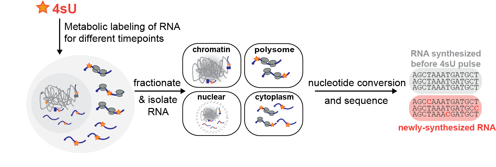
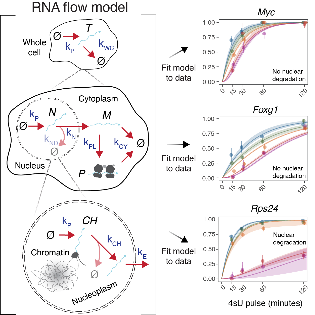
Using subcellular TimeLapse-seq, we tracked the age of RNAs as they moved across subcellular compartments and determined RNA half-lives at subcellular resolution using a Bayesian modeling framework. This measured the rates at which transcripts were released from chromatin, exported from the nucleus, and loaded onto polysomes for all human mRNAs, as well as nuclear and cytoplasmic degradation rates.
A simple RNA life cycle model did not fit our data for ~10% of genes; instead, a model including substantial nuclear mRNA degradation was required. Hundreds of genes were predicted to undergo nuclear degradation (PUNDs), and more than 85% of the transcripts encoded by these genes are never exported from the nucleus. Many PUND genes participate in gene expression and RNA biology, including ribosome subunits, RNA splicing and nuclear mRNA export factors. PUND transcripts possess features such as incomplete splicing and long poly(A) tails, which may aid their identification by nuclear degradation pathways.
Open questions
How can RNA dynamics be measured in any RNA-seq experiment?
Metabolic labeling separates synthesis from decay, but the chemical recoding step it usually requires damages RNA and does not survive droplet-based single-cell protocols. Analog intrinsic recoding sequencing (AIR-seq) removes that step. Cells are labeled with N4-hydroxycytidine, the active compound of the antiviral molnupiravir, which is incorporated into RNA by RNA polymerase II and base-pairs ambiguously, so new transcripts carry C-to-T and T-to-C mismatches in otherwise standard libraries.
Fraction-new estimates from AIR-seq agree with subcellular TimeLapse-seq, hold across library types and sequencing depths, and work in single cells with the unmodified 10x workflow. A single labeled snapshot orders cells around the cell cycle and gives new RNA a roughly one-hour lead over total RNA, which sharpens RNA velocity estimates. Modeling those rates revealed offset synthesis and degradation programs for nuclear- and mitochondrial-encoded OXPHOS transcripts that leave steady-state abundance unchanged.
Open questions
Background reading: Hansen & Couvillion et al., bioRxiv 2026
Method
Measuring RNA flow across subcellular compartments
Using subcellular TimeLapse-seq to measure the rates of transitions between cellular compartments, we found that RNAs do not flow across the cell at the same rates. Our study revealed wide variations (>100-fold) in RNA flow rates between genes and subcellular compartments. Functionally related genes experienced similar rates of RNA flow, and the targets of many RNA binding proteins exhibited different flow rates compared to other genes.
We measured poly(A) tails with subcellular resolution and showed that tail lengths reflect subcellular RNA half-lives. Machine learning identified genetic and molecular features that predict RNA flow, including transcription factors and sequence elements.
Open questions
Background reading: Ietswaart et al., Mol Cell 2024
Program two
Mitonuclear co-regulation
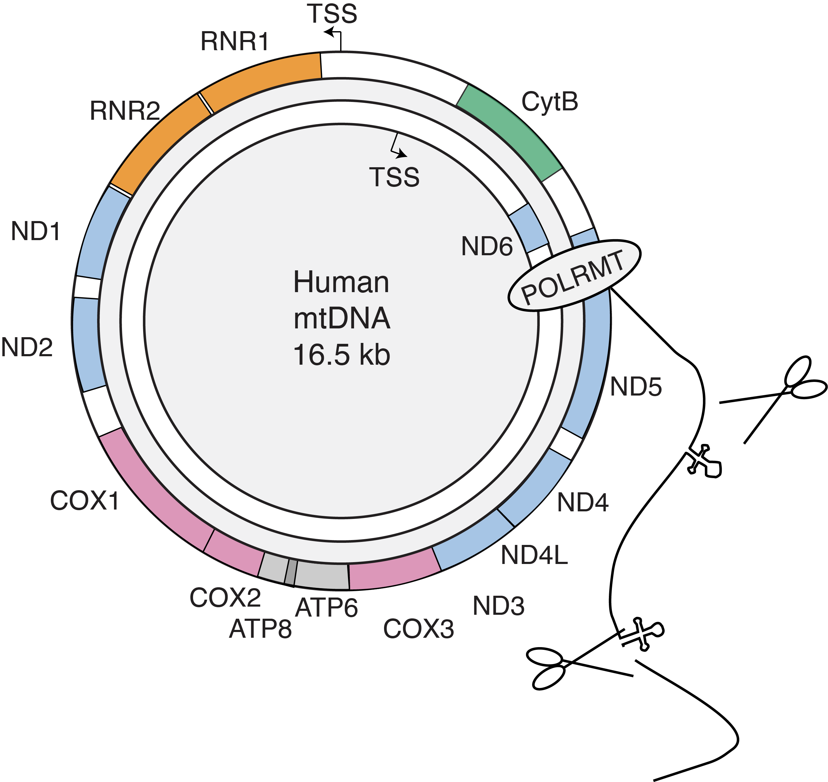
Human mtDNA, originating from a eubacterial genome, has significantly reduced in size due to gene loss or transfer. The remaining 16.5 kb genome encodes 13 proteins, all core subunits of the OXPHOS complexes, while the other subunits are nuclear-encoded. How eukaryotic cells coordinate gene expression between two disparate genomes in separate cellular compartments, using separate machinery and regulatory processes, is the question this program addresses.
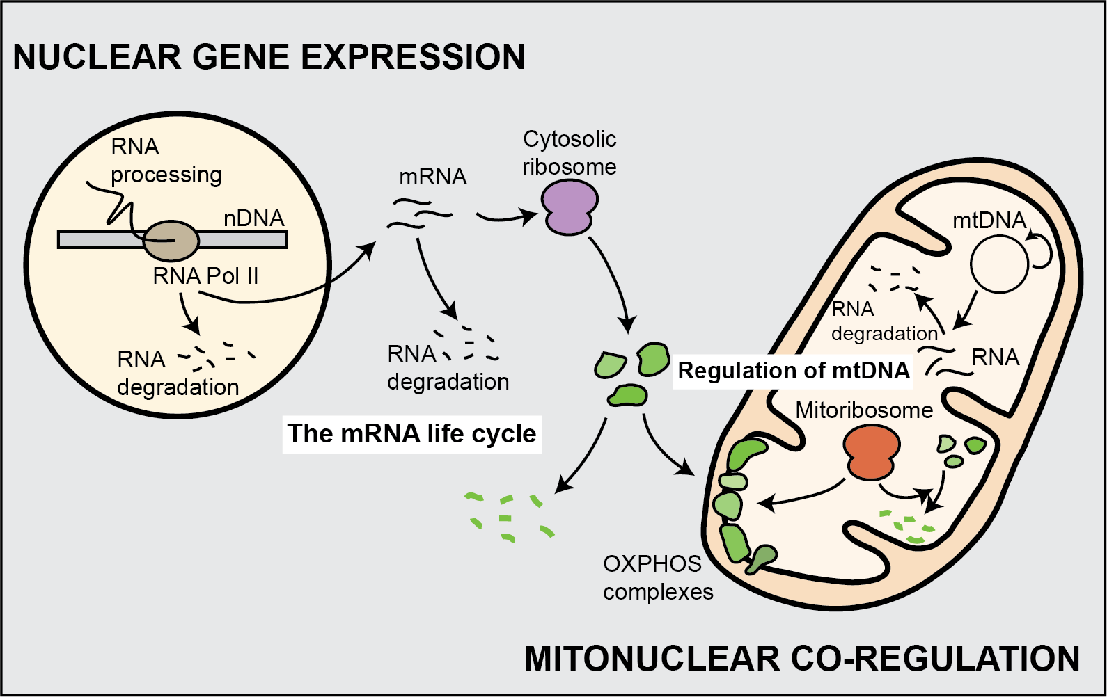
Our first studies in this area asked whether nuclear and mitochondrial gene expression processes were coordinated. By adapting ribosome profiling to assess mitochondrial protein synthesis, we demonstrated that the translation of nuclear- and mitochondrial-encoded OXPHOS subunits is balanced in both yeast and human cells. To pinpoint the stages of mitochondrial gene expression that are most regulated, we are now quantitatively dissecting each step.
The mitochondrial mRNA life cycle. The non-coding region of the genome contains two promoters that generate near-genome-length polycistronic transcripts, which are then cleaved into individual mRNAs with little to no 5′ UTR. We measured RNA production, processing, ribosome arrival and turnover using TimeLapse-seq and direct nanopore RNA sequencing. By modeling protein synthesis levels from these data, we found that mitochondrial mRNA turnover rates and translation efficiency mainly account for protein synthesis levels in human mitochondria.
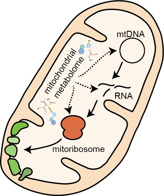
Which nuclear-encoded factors and metabolites control mitochondrial gene expression?
Discovered by genetic screens, yeast translation activators are nuclear-encoded and imported into mitochondria to promote the translation of specific mRNAs via their 5′ UTRs. To identify human genes that control mitochondrial gene expression, we developed genome-wide CRISPR screen strategies to identify factors that affect the balance of Complex IV subunits, mitochondrial-encoded COX1 and nuclear-encoded COX4. We identified known regulators such as TACO1, whose mutations cause Leigh syndrome, alongside genes with unclear mitochondrial functions. Among the latter, we are initially focusing on PREPL, a protein highly expressed in the brain, and NME6, a member of the nucleotide diphosphate kinase (NDPK) family.
How does mitonuclear co-regulation work in neurons?
Neurons are highly dependent on mitochondrial function, and their complex, polarized morphology offers a challenge for balanced nuclear and mitochondrial gene expression. We aim to determine whether this structure makes neurons more susceptible to mitonuclear imbalance due to genetic or physiological stress, and whether this is relevant to mitochondrial malfunction in neurodegenerative disease.
Open questions
Background reading: Couvillion et al., Nature 2016 · Soto et al., Genome Biol 2022 · Kramer & Prakash et al., Nat Cell Biol 2023 · Isaac et al., Nat Struct Mol Biol 2024 · McShane et al., Mol Cell 2024
Method
Single-molecule views of mitochondrial DNA packaging
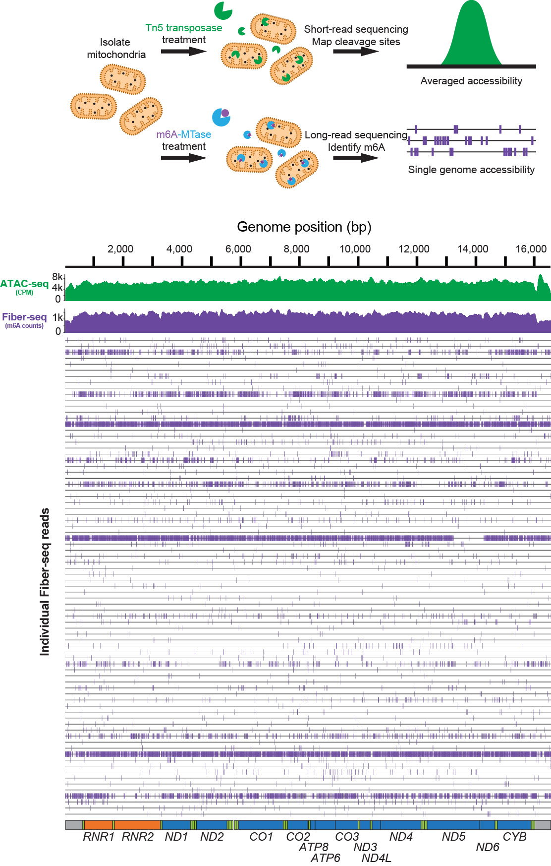
Human cells contain hundreds to thousands of mtDNA molecules distributed throughout the mitochondrial network. Each molecule, more than 5 µm in length, is compressed into a nucleoprotein complex called a nucleoid. To measure how individual nucleoids are packaged, we developed mtFiber-seq, a long-read single-molecule accessibility method that profiles full-length mtDNA molecules at near single-nucleotide resolution.
mtFiber-seq revealed that human mtDNA undergoes largely all-or-none global compaction, with most nucleoids existing in an inaccessible, inactive state. Highly accessible nucleoids are occupied by transcription and replication machinery and selectively form the triple-stranded displacement loop. We also found that TFAM controls the fraction of inaccessible nucleoids through a nucleation-and-spreading mechanism, identifying genome accessibility as a layer of mitochondrial gene regulation.
Isaac et al., Nature Structural & Molecular Biology 2024